
This is one of the main muscles in the calves. It mainly serves as an attachment point for the muscles of the lower leg.

It acts as the main weight-bearing bone of the leg. Also called the shin bone, the tibia is the longer of the two bones in the lower leg. This area is commonly referred to as the calf. The lower leg extends from the knee to the ankle. In addition, they help evenly distribute weight, providing balance and stability. They’re discs of cartilage that act as shock absorbers. The knee contains two menisci (plural), known as the medial meniscus and lateral meniscus. They help reduce friction and inflammation in the knee. Bursae (plural) are small sacs filled with fluid in the knee joint. Some of the most important structures include: The knee contains a variety of structures that help it support weight and allow a range of movements. What’s the difference between tendons and ligaments? Find out here. The quadriceps tendon attaches the quadriceps muscle to the patella. The largest tendon in the knee is the patellar tendon. They’re found on the ends of muscles, where they help attach muscle to bone. Tendons are also bands of connective tissue. This provides stability for the inner knee. This prevents the knee from moving too far backward. This prevents the tibia in the lower leg from moving too far forward. They help support joints and keep them from moving too much. Ligaments are bands of connective tissue that surround a joint. Also called the kneecap, the patella serves as a point of attachment for different tendons and ligaments. It also allows for rotation and pivoting. In addition to bearing the weight of the upper body, the knee allows for walking, running, and jumping. The knee joins the upper leg and the lower leg. The adductors are five muscles located on the inside of the thigh. Try these three quadriceps stretches if you’re a runner. They allow the knees to straighten from a bent position. The quadriceps are four muscles located on the front of the thigh.
#COMPLETE ANATOMY OF HIP JOINT HOW TO#
Learn how to prevent and treat hamstring pain. The hamstrings are three muscles located on the back of the thigh. It can account for about a quarter of someone’s height. Also called the thigh bone, this is the longest bone in the body. It’s the area that runs from the hip to the knee in each leg.

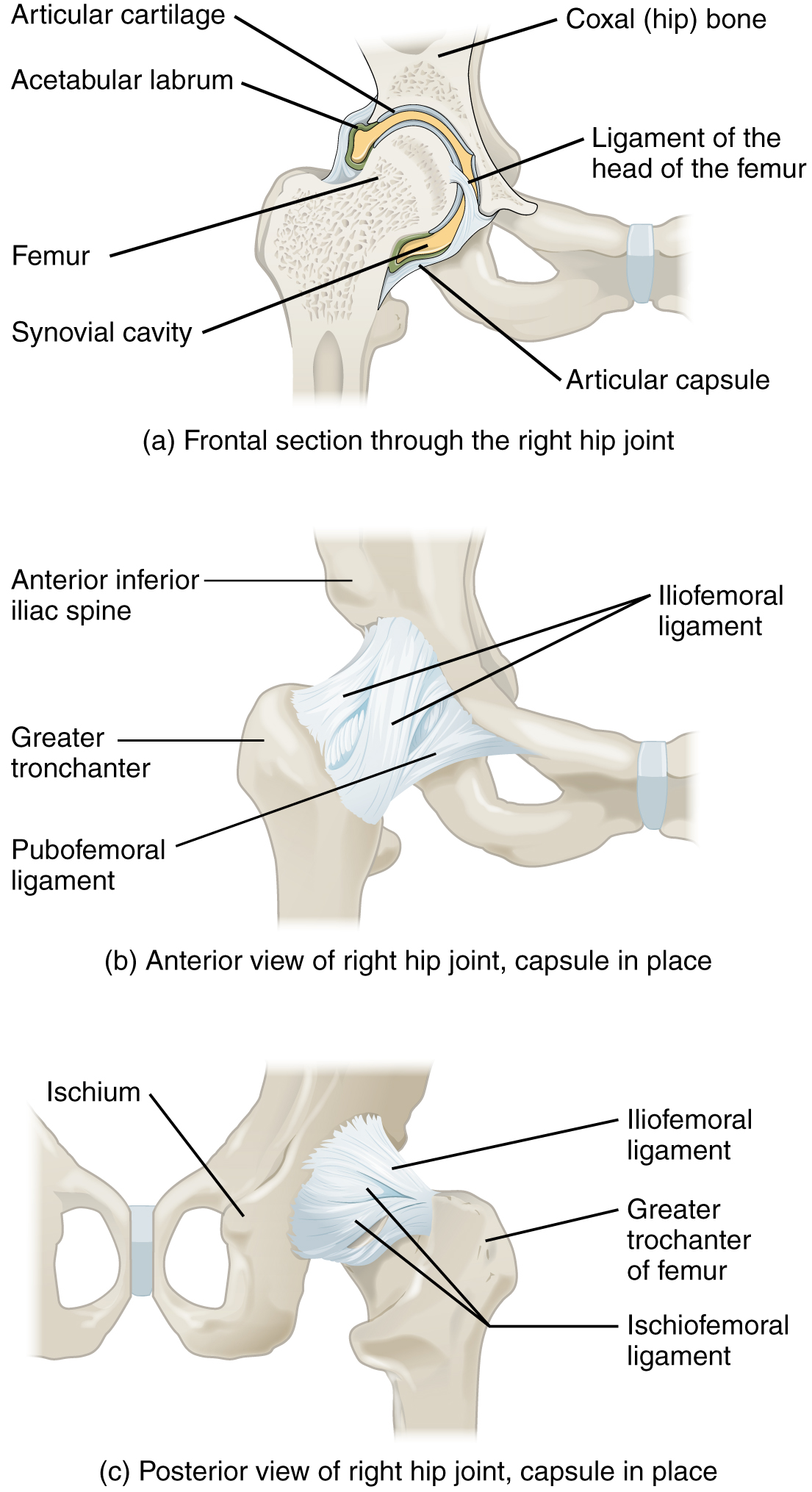
Labrum tears are not an uncommon hip injury. The labrum is a circular layer of cartilage which surrounds the outer part of the acetabulum effectively making the socket deeper to provide more stability for the joint. Ischiofemoral ligament, which attaches to the ischium (the lowest part of the pelvis) and between the two trochanters of the femur.Pubofemoral ligament, which attaches the most forward part of the pelvis known as the pubis to the femur.Iliofemoral ligament, which connects the pelvis to the femur at the front of the joint.The most notable ligaments in the hip joint are: LigamentsĪs noted above, the stability of the hip joint is directly related to its muscles and ligaments. This membrane nourishes and lubricates the joint. Inside the capsule, the surfaces of the hip joint are covered by a thin tissue called the synovial membrane. The joint capsule is a thick ligamentous structure surrounding the entire joint. You may hear your hip surgeon refer to the capsule or socket, when describing the structure of the hip joint. Hip capsule (socket) illustration (image courtesy of: Smith & Nephew)


 0 kommentar(er)
